HOW TO TREAT BED SORE AND THEIR MANAGEMENT SURGICAL ASPECT
Types of Bedsores (Pressure Ulcers)
Bedsores, also known as pressure ulcers, can be categorized into different stages based on their severity:
- Stage 1: In this stage, the skin is intact but may appear red or discolored. It may also feel warm or firm to the touch. In individuals with darker skin tones, the area may appear blue or purple.
- Stage 2: The skin is partially damaged, presenting as an open wound, blister, or shallow ulcer. The area may be red, pink, or swollen, and there may be some drainage or pus.
- Stage 3: At this stage, the ulcer extends into the deeper layers of the skin, causing a deep crater-like wound. Fat may be visible in the ulcer, and there may be signs of infection such as pus or a foul odor.
- Stage 4: The most severe stage, where the ulcer extends through all layers of the skin, affecting muscle, tendons, and even bone. The wound may appear blackened or necrotic, and there is a high risk of infection and complications.
- Unstageable: In addition to these stages, there are also unstageable bedsores, where the extent of tissue damage is obscured by eschar (dead tissue) or slough (yellow or white tissue). These wounds require debridement to remove the non-viable tissue before staging can be determined.
Each stage requires specific treatment and management, so it's crucial to accurately assess and classify bedsores to provide appropriate care. Regular monitoring and prevention strategies are essential to avoid the development of bedsores in vulnerable individuals.
How to Bed Sore Caring
Caring for bedsores (pressure ulcers) in older patients involves several steps:
- Relieve Pressure: Ensure the patient is repositioned frequently to relieve pressure on the affected area.
- Cleanse the Wound: Gently clean the wound with mild soap and water, or a saline solution, to remove any debris or bacteria.
- Apply Dressings: Use appropriate dressings recommended by a healthcare professional to protect the wound and promote healing.
- Monitor for Infection: Watch for signs of infection such as increased redness, warmth, swelling, or drainage from the wound. Consult a healthcare provider if infection is suspected.
- Nutrition and Hydration: Ensure the patient is well-nourished and hydrated, as proper nutrition is crucial for wound healing.
- Pain Management: Manage pain with medications or other interventions as prescribed by a healthcare provider.
- Medical Attention: Seek medical attention if the wound does not show signs of improvement or if it worsens despite proper care.
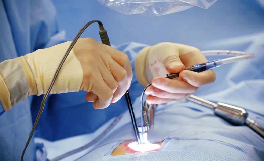
Surgical View of Bed Sore
When surgeons approach bedsores (pressure ulcers), they typically view them as complex wounds that require careful evaluation and tailored treatment plans. Here are the key surgical perspectives on bedsores:
- Assessment: Surgeons assess the severity and depth of the wound, as well as any signs of infection or necrosis. This evaluation helps determine the appropriate surgical approach and interventions needed for optimal healing.
- Debridement: Surgical debridement involves removing necrotic (dead) tissue, slough, and debris from the wound. This step is crucial for promoting the growth of healthy tissue and preventing infection. Debridement techniques may include sharp debridement (using surgical instruments), enzymatic debridement (using topical enzymes), or mechanical debridement (such as wet-to-dry dressings).
- Flap Reconstruction: In cases of extensive tissue loss or deep wounds, surgeons may perform flap reconstruction. This involves transferring healthy tissue from nearby areas of the body to cover the wound. Flap reconstruction helps restore tissue integrity, promote wound healing, and reduce the risk of recurrence.
- Skin Grafting: Surgeons may also use skin grafts to cover large or deep wounds. Skin grafting involves taking skin from a healthy donor site (autograft) or a cadaver (allograft) and transplanting it onto the wound site. This procedure helps accelerate wound healing and improve the cosmetic appearance of the affected area.
- Negative Pressure Wound Therapy (NPWT): After surgical intervention, surgeons may utilize NPWT to facilitate wound healing. NPWT involves applying a vacuum dressing over the wound, which helps remove excess fluid, reduce swelling, and promote the formation of granulation tissue.
- Postoperative Care: Surgeons provide comprehensive postoperative care, including wound monitoring, dressing changes, pain management, and infection prevention. Patients may also receive instructions on proper positioning, pressure relief techniques, and nutrition to support wound healing.